Focus on precision medical catheters
language:  ∷
∷ 
∷ ∷ 
The incidence of peripheral nerve injury is 1.2%~2.8% in trauma patients. Peripheral nerve injury can lead to the loss of neuronal communication between sensory and motor nerves between the central nervous system and the surrounding organs, and seriously affect the daily activities of patients. The current preferred treatment is tension-free suture of the epineurium via microsurgery, and autogenous nerve graft is the gold standard in cases where the nerve defect of end-to-end suture cannot be achieved. However, autologous nerve transplantation has a limited donor nerve source and a long operation time, which is easy to be hindered by the morbidity and loss of function of the donor site. Therefore, the treatment effect of peripheral nerve injury is not ideal. The repair and regeneration of peripheral nerves remains one of the greatest challenges in tissue engineering and medicine.
Therefore, the author chose artificial nerve ducts as axon guiding channels. These tubular structures can connect the Spaces of severed nerves to promote the reconnection of neurons. Nerve ducts can act as a physical barrier to the external environment and a physical guide for regenerated axons to pass through the gap injury. At the same time, they can retain the naturally released nerve growth factors secreted by the damaged nerve stump and reduce the invasion of fibroblasts from scar tissue to the injured site. At present, progressive progress has been made in the development of nerve conduits. This paper reviews the design progress of nerve conduits for bridging segmental nerve defects, and reports as follows.
Ideal properties of artificial nerve conduit
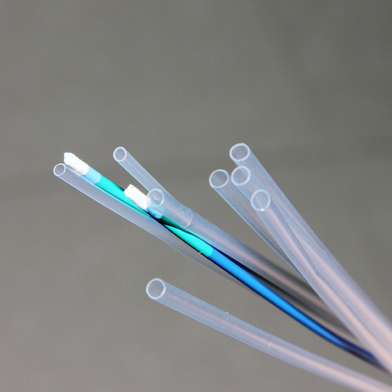
In order to provide an ideal channel for axon regeneration, artificial nerve conduit materials should have the following physical properties: (1) permeability. Before vascularization of the conduit, nutrients and oxygen need to be diffused to the repair site after peripheral nerve injury to ensure the activity of supporting cells; (2) Adaptability, artificial nerve ducts should have good histocompatibility to avoid mechanical damage to surrounding tissues and axon regeneration; (3) Swelling, local swelling may block the pipeline, hinder the regeneration of nerves in the pipeline, or directly damage the regeneration of nerves in the pipeline; (4) Degradation rate, the ideal artificial nerve duct should remain intact until the axon reinnervates the distal nerve pathway from the stump through the space, and then gradually degrades. If the degradation rate is too fast, it may lead to swelling and local inflammation. If the degradation rate is too slow, the catheter may compress the nerve, leading to chronic immune rejection.
The main material of artificial nerve conduit
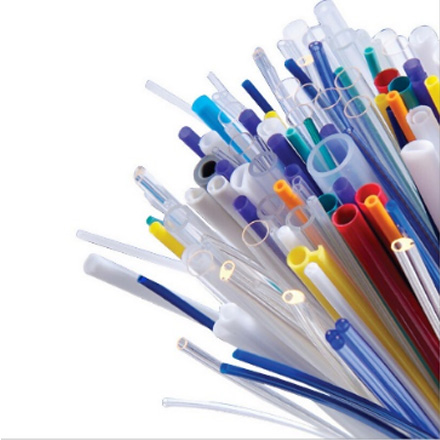
The use of artificial nerve catheters can be traced back to 1989, when Merle et al. used silicone tubes to bridge the defective nerve sites. Subsequently, scholars demonstrated the feasibility of silicone catheters for nerve repair. However, the inabsorbability of silica gel results in permanent wrapping of the implant, which is accompanied by chronic nerve compression, resulting in blocked nerve regeneration and often requiring a second surgical intervention. These defects limited the use of silica gel catheters and facilitated the development of absorbable synthetic catheters. Since the early 1990s, numerous experimental studies have led to the development of a variety of artificial nerve conduits.
Polyglycolic acid Polyglycolic acid is a biodegradable rigid thermoplastic polyester with very low solubility in organic solvents. As a kind of suture material commonly used in clinic. Silicone tubes were the first material used to construct nerve ducts when their limitations were realized.
Polyglycolic acid has excellent mechanical properties and can be rapidly degraded into lactic acid within 3 months. Polyglycolic acid is the first synthetic, highly porous, bioabsorbable nerve conduit approved by the U.S. Food and Drug Administration, although it may be degraded before peripheral nerve regeneration and its lactate degradation products may be toxic to peripheral nerve regeneration. Mackinnon et al. used polyglycolic acid catheter to perform 15 secondary reconstruction operations for digital nerve defects within 3cm, and most of the patients had good prognosis. Rosson et al. bridged the median nerve defect of 15 to 40mm, and the patient's motor function recovered well after surgery.
Some scholars have extended the use of polyglycolic acid catheters to a wider range of motor nerve defects. We⁃ber et al. compared the use of a polyglycolic acid catheter for peripheral nerve injury repair with standard surgery and found that the use of a polyglycolic acid catheter for nerve defect repair within 4mm was more effective than end-to-end repair.
Plla can be made from lactic acid extracted from corn, beet or wheat, and has good biocompatibility. Therefore, PLLA is used as nerve conduit material in many biological studies. In the rat model of sciatic nerve injury, by creating multilayer polylactic acid nerve ducts to obtain sufficient mechanical strength at the site of nerve injury, successful regeneration of peripheral nerves through the defect was observed 8 weeks after surgery. Some scholars made a PVC nerve conduit in the rabbit sciatic nerve transection model by soaking precipitation method to bridge a 20mm defect. These conduits have large pores on the outside and interconnected micropores in the inner layer to provide a higher outflow rate than the inflow rate. According to electrophysiological and behavioral analysis, the nerve function recovered 80% after 18 months.
However, the pure polylactic acid nerve duct is prone to deformation and collapse, and the degraded product is acidic, which may stimulate the surrounding tissues to produce inflammatory reactions and hinder the repair and regeneration of peripheral nerves. Therefore, it is necessary to work together with other materials. The rat sciatic nerve defect was treated by polylactic acid combined with glycolic acid copolymer. After 3 months, the nerve injury site was observed by electron microscopy. The results showed that there were more myelin fibers around the lesion, the myelin sheath was complete and arranged regularly, and the myelin sheath was closely adjacent to the axon membrane.
The polycaprolactone nerve catheter is the latest synthetic catheter approved by the US Food and Drug Administration. It has the advantage that it degrades to produce fewer acidic products and therefore is theoretically less toxic to surrounding tissues. However, the rigidity of the polycaprolactone nerve catheter makes it more difficult to operate in clinical use. Pego et al. studied the physical and chemical properties of Sanya methyl carbonate and caprolactone copolymer to evaluate its potential in adaptability and slow degradation ability. They believed that Sanya methyl carbonate and polycaprolactone copolymer with high caprolactone content had good physical properties and was suitable for the preparation of porous artificial nerve conduit.
The experimental results showed that the 1cm defect of sciatic nerve in rats was repaired by using the polycaprolactone nerve catheter. After 18 weeks, the peripheral nerve axons grew well and the sensory and motor functions were obviously restored. In a prospective study by Bertleff et al., in three groups stratified by space length, the prognosis for end-to-end repair was comparable to that of standard surgery. However, in 28 peripheral nerve repair operations performed with a Pcaprolactone catheter, only 25% had a good prognosis and 35% had complications. Due to these conflicting results, the current efficacy of polycaprolactone catheters remains unclear.
Collagen is a family of 26 proteins with an extended rod-like triple helix structure that is a major component of the extracellular matrix. Type I collagen is one of the oldest natural polymers used as biomaterials, accounting for about 30% of mammalian musculoskeletal tissue. Collagen is thought to promote cell adhesion and survival, and promote cell proliferation. However, it takes 48 months for collagen ducts to degrade, and this relatively long degradation time may bring the risk of fibrosis and nerve compression.
Still, a nerve catheter made of type I collagen has been approved by the U.S. Food and Drug Administration. As a collagen-based semi-permeable wrapping material, it can unfold and self-curl to maximize the match of damaged tissue; In addition, the structure of the semi-permeable membrane allows the diffusion of nutrients while preventing the migration of fibroblasts. Subsequently, the approved type I collagen nerve catheter was used in the clinic.
Keilhoff et al. used the rat sciatic nerve model to test the ability of type Ⅰ collagen nerve conduit as a potential nerve guiding substrate, and believed that type Ⅰ collagen could be used as a template to design "living" nerve conduit, which might be able to ensure nerve regeneration by extending the gap between nerve defects. In a retrospective study, a collagen nerve catheter was used to repair sensory nerve defects in the hand within 20mm, and the patients had good recovery results. At the same time, a retrospective study was conducted on neuroma patients who underwent collagen nerve catheter repair. Among the 11 patients who received the treatment, 5 cases recovered well and had normal function. Taras et al. used collagen nerve catheter to repair isolated digital nerve laceration lesions ranging from 5mm to 15mm, and the recovery rate reached 73%. These results suggest that collagen nerve catheters are safe and effective for peripheral nerve defect lesions within 20mm.
Fibrin, a glycoprotein linked by disulfide bonds, is one of the main proteins of the extracellular matrix and can interact with collagen, heparin, fibrin and cell surface receptors, playing an important role in cell adhesion, migration and differentiation. Fibrin can accelerate angiogenesis, increase the number of leukocytes around the lesion and promote the proliferation of macrophages. When used as a suture repair material, the solidified semi-solid fibrin hydrogel effectively stops bleeding and enhances the integrity of the repair.
In addition, animal studies have shown that the use of fibrin for end-to-end nerve repair can reduce the incidence of complications such as inflammation and fibrosis compared with suture repair. Moshahebi et al. added fibrin to alginate, which confirmed that fibrin can accelerate the growth of peripheral nerve axons by affecting the activity of Schwann cells. Despite good progress in a large number of experimental studies, clinicians remain cautious about using fibrin for nerve repair, in part due to inconsistent reports on the tensile strength of repair sites and concerns that fibrin may inhibit nerve regeneration.
Meanwhile, the use of collagen catheter filled with fibrin to repair the 10mm rat sciatic nerve defect also confirmed that fibrin can promote the repair and regeneration of peripheral nerves, suggesting that fibrin can be used as an adjuvant for peripheral nerve repair. However, the effect of fibrin in promoting peripheral nerve regeneration needs to be further studied clinically.
Chitin and chitosan Chitin, a component of the cytoskeleton in insect and crustacean exoskeletons, is a major source of chitosan and the second most abundant polysaccharide on Earth after starch. Chitosan is a linear polysaccharide consisting of glucosamine and n-acetylglucosamine units connected by glycosidic bonds. Chitosan is the deacetylated form of chitin. Due to its good biocompatibility, biodegradability and bioactivity, chitosan is considered as a good biomaterial and has a wide range of biomedical applications. Its affinity for nerve cells and its ability to promote survival and axon growth in vitro suggest that chitosan can be used as a scaffold for peripheral nerve axon regeneration.
Zheng et al. used chitosan nerve catheter combined with bone marrow mesenchymal stem cells to promote peripheral nerve regeneration, and found that bone marrow mesenchymal stem cells could differentiate into neural stem cells in rats, which could effectively repair 8mm long peripheral nerve defects after differentiation. However, the use of chitosan nerve catheter still has some limitations, such as high degradation rate and poor mechanical stability.
Silk fibroin is a hydrophobic domain containing short chain amino acids synthesized by special epithelial cells in the glands of mites, butterflies and moth worms. The strength of silk fibroin comes from the size of the interval between these hydrophobic domains and hydrophilic fragments. Compared with other protein-based biomaterials, silk fibroin has many advantages, such as relatively low risk of infection and less repulsive to other materials; The high biocompatibility and biodegradability of silk fibroin are another advantage worth considering in tissue engineering applications.
The nerve catheter synthesized from silk fibroin proved to be an ideal material to heal the 13mm peripheral nerve injury defect within 12 weeks. Gu et al. used an artificial nerve catheter made of silk fibroin and chitosan and implanted with Schwann cells to repair peripheral nerve defects. Morphological and electrophysiological analysis showed that the regeneration effect of the nerve conduit was close to that of autologous nerve transplantation, indicating that the nerve conduit had a good ability to promote the repair and regeneration of peripheral nerve injury. Therefore, silk fibroin is expected to be an ideal artificial nerve conduit material for repairing peripheral nerve injury.
Gelatin Gelatin is a low cytotoxic crosslinking agent that is widely used in tissue engineering after being cross-linked with various chemicals. Gamez et al. implanted gelatin nerve catheter into the lesion of right sciatic nerve defect in adult male rats for 1 year and observed significant recovery of sciatic nerve function, morphology and electrophysiological response. Liu et al. prepared a proanthocyanidin cross-linked gelatin nerve catheter with rough outer surface, and applied the catheter to the rat sciatic nerve injury model of 10mm. After 8 weeks, the regenerated nerve fibers could be seen passing through and beyond the defect area.
Nie et al. implanted gelatin and chitosan nerve ducts containing transforming growth factor β1 into the 10mm defect lesion of rat sciatic nerve, and the inner surface of the ducts remained intact during the regeneration process, which could prevent the inward growth of connective tissue. Functional recovery, electrophysiological testing, and immunohistochemical analysis showed that nerve conduction velocity, regenerated myelin sheath area, and myelinated axon count were similar to those in the autograft group. Many experimental results show that gelatin as artificial nerve conduit material can effectively promote the repair and regeneration of peripheral nerve injury
Keratin Keratin is a protein produced by keratinocytes. The cysteine in this structure is rich in sulfur and plays an important role in the bonding of hair. Hair fibers are mainly composed of cross-linked keratin, from which free keratin molecules can be extracted for the construction of biological materials. Keratin forms hydrogels by oxidized keratin crosslinking. Compared with autologous nerve grafts, keratin biomaterial has nerve induction ability. In a rat model of 15mm sciatic nerve defect, a catheter filled with keratin gel was used. After treatment, Schwann cells and axons grew actively in the nerve defect area.
Pace et al. used keratin hydrogel as the lumen filling of nerve conduit for the model of median nerve transverse injury in macaques. After 12 months of treatment, electrophysiological results showed that keratin nerve conduit could promote the repair and regeneration of peripheral nerve injury. Nerve catheters have shown ideal effects in many peripheral nerve injury lesions in animal experiments, but the effect of artificial nerve catheters on a large range of peripheral nerve defects in multiple parts of the human body still needs to be confirmed by further studies. There are studies using collagen and polyglycolate nerve catheter to repair brachial plexus, median nerve and ulnar nerve injury, the patients did not recover motor and sensory function, the prognosis is not ideal. Reexamination of the repaired site revealed that a significant neuroma had formed outside the repaired site, requiring resection of the neuroma and repair with an autologous nerve graft.
In fact, many cases of artificial nerve catheter repair of large nerve defects involve the use of a smaller diameter catheter to bridge the defective nerve stump and the combination of the nerve catheter with a smaller autologous nerve graft. On the other hand, although promising results have been obtained from the data collected in experimental animal studies, we should be cautious in clinical applications because animals can achieve tissue morphology and functional recovery in a mode of injury that humans cannot overcome. For example, a rodent nerve can regenerate spontaneously within five months through a 4.5-meter nerve defect, while a completely severed nerve in a human has to be surgically repaired before functional recovery can be achieved. With the continuous improvement of biological level and the continuous breakthrough of tissue engineering and manufacturing technology, the design of artificial nerve conduit is constantly undergoing revolutionary changes.
Biodegradable nanomaterials hold promise for the manufacture of novel nerve conduits, at the right density and three
 WeChat scan code

support hotline0755-23251960

mobile phone191 7359 3045
Copyright © SOKO Medical All Rights Reserved. Add:Shenzhen,CHINA 粤ICP备2022121184号  粤公网安备 44030402005873号